Digital Imaging is the new technology that allows dentists to accurately record facial and dental images into digital information and to be able to analyze, diagnose, measure and treat dental conditions. The standardization of these records allows for digital files to be transferred and archived without the loss of quality and coordination with specialists and referring doctors in dramatically improved. The ability to be able to refer back to a photograph or radiograph that may have been recorded in prior years, can help the doctor find and determine the diagnosis, severity and susceptibility of any area of concern and influence the treatment that is best for an individual. The radiographic technology utilize significantly lower exposures of radiation and create an image that is far more versatile and with higher image fidelity resolution.
The dentist can view the images on the computer monitor and modify the images with software that allows the doctor to diagnose and discover problems at their earliest stages. These images can be shared within a team of doctors that can facilitate virtual consultations to insure the best possible patient care. Most recently, with the advent of cone beam computer tomographic scan imaging, or CBCT scans, an image can be obtained, manipulated and created into an incredible 3 dimensional image of the skull that can be used in the treatment planning and placement of dental implants with accuracy and predictability that was never possible before.
One of our most important practice goals is staying on the cutting edge of medical and dental technology. This is why we are proud of using the “gold standard” in dental and facial imaging of hard tissues and diagnostic study, Cone Beam CT scan: Here is a brief introduction.
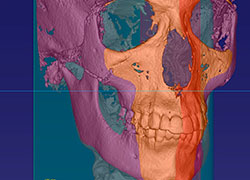
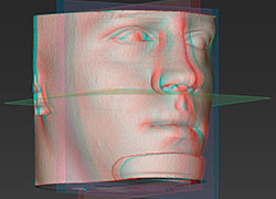
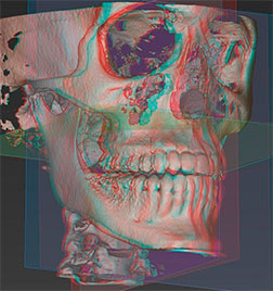
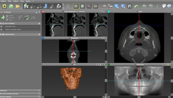
Traditional dental X-rays while certainly effective for detecting cavities (caries) and other minor irregularities have definite limitations when it comes to more in-depth dental and maxillofacial surgery procedures. This is where Cone Beam CT technology comes in. Projecting X-rays in a distinct cone-shaped beam and using technology similar to that of a traditional medical CAT scan, Cone Beam CT renders a clear, 3-D image of the teeth, facial - jaw bones, vital structures and more.
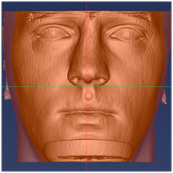
Three dimensional (3-D) imaging provides a complete view of the upper and lower jaw bones, providing the most thorough diagnostic information possible of any type of radiographic examination. This radiographic modality allows doctors to more accurately diagnose and treat patients more predictably than with conventional two dimensional (2D) imaging. Every aspect of the facial skeleton can be evaluated with distortion-free images reconstructed from true data gathered by an advanced digital sensor. Any measurements taken from any image in the scan will always have a one-to-one ratio, and therefore have no magnification error. In addition, the scan can be used to develop extremely accurate 3-D reconstructions of the face and head, with the ability to view multiple images in any dimension with slices as small as 0.2mm in thickness. Moreover, the software allows the surgeon to measure relative bone densities in any area of any image obtained.
Here are a few ways Cone Beam CT is used today: